

News • Nanometer-scale imaging
Microscopy technique enables 3D super-resolution
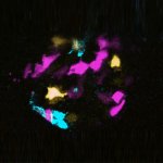
Research team led by Göttingen University combine two techniques to achieve isotropic super-resolution imaging.


Research team led by Göttingen University combine two techniques to achieve isotropic super-resolution imaging.
To develop new drugs, detailed knowledge about nature’s smallest biological building blocks is required. A new microscopy technique that allows proteins, DNA and other tiny biological particles to be studied in their natural state.
Researchers from Boston University School of Medicine have developed a novel artificial intelligence algorithm to assess digital pathology data.
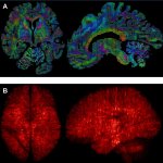
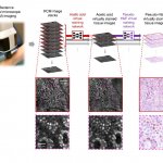
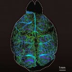
A team led by scientists from Amsterdam have combined MRI and microscopy to produce 3D images of two entire brains with a previously unmatched level of detail.
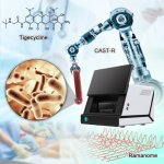
When treating acute infections, health care providers must quickly identify the best antibiotics for fighting the infection. An automated system provides swift, accurate results for determining the best antibiotics at the right dose.
MediSCAPE, a high-speed 3D microscope, can see real-time cellular detail in living tissues to guide surgery, speed up tissue analyses, and improve treatments.

The team at the Institute of Medical Device Technology at the University of Stuttgart, Germany, is developing methods to produce top-quality medical devices at affordable prices. Professor Dr Peter Pott, the director of the institute, turns to 3D printers to successfully realize his vision of “high end at low cost”.
Ever since the Abbe diffraction limit of conventional microscopy has been surpassed, super-resolution techniques have been diving ever deeper into the most miniscule details of molecular structures. We spoke with Prof. Dominic Zerulla, whose company PEARlabs is developing an imaging technique that sets out to push the boundaries once more – by looking at in-vivo nano-scale processes in motion.
Researchers at the McKelvey School of Engineering at Washington University in St. Louis found a way to significantly reduce the noise and maintain image quality while reducing the laser energy needed to generate images by 80%.
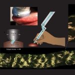
A 'new technology shows promise by analyzing images of suspicious-looking lesions and quickly producing a detailed, microscopic image of the skin, bypassing several standard steps typically used for diagnosis - including skin biopsy, tissue fixation, processing, sectioning and histochemical staining.
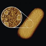
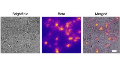
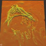
The sharpest images ever of living bacteria have been recorded by researchers at University College London, revealing the complex architecture of the protective layer that surrounds many bacteria and makes them harder to be killed by antibiotics.
If the suspicion of Alzheimer's disease creeps up, those affected must prepare themselves for lengthy and complex procedures until the case is clear. A team from Empa and the Cantonal Hospital of St. Gallen is now in the process of developing a blood test that will enable a reliable diagnosis using atomic force microscopy (AFM).
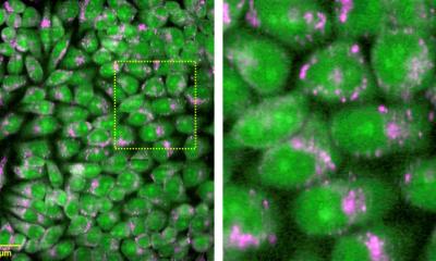
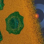
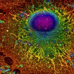
Researchers at Umeå University now demonstrate a method by which specific cell types in human organs can be studied with micrometer precision. The method can be used to reveal previously unrecognised alterations in the pancreas, but it can also be used to study other human organs and diseases.
Scientists use super-resolution microscopy to study previously undiscovered cellular worlds, revealing nanometer-scale details inside cells. This method revolutionized light microscopy and earned its inventors the 2014 Nobel Prize in Chemistry. In an international collaboration, AI researchers from Tübingen have now developed an algorithm that significantly accelerates this technology.
Researchers from ETH Zurich and University of Zurich have developed a new microscopy technique that lights up the brain with high resolution imagery. This allows neuroscientists to study brain functions and ailments more closely and non-invasively.

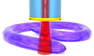
University of Birmingham scientists have developed a new microscopic imaging approach to take a closer look at 3D-printing for developing future patient implants, as well as improved disease modelling and drug screening. Additive manufacturing (3D printing) platforms create bioprinted structures by moving a special bioink, containing cells, biomolecules and materials, through a narrow tube, but…
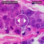
Did you know that changing microscopy camera can make a real difference to your microscopy workflow? Packed full of smart features to make your imaging faster and easier, Olympus’ new DP23 and DP28 (4K) cameras enable streamlined, comfortable sample analyses at the monitor and easier, smoother conferencing. Get the most out of the monitor view and discover the many benefits of the DP23 and DP28…
Using an ordinary light microscope, engineers at the Massachusetts Institute of Technology (MIT) have devised a technique for imaging biological samples with accuracy at the scale of 10 nanometers — which should enable them to image viruses and potentially even single biomolecules, the researchers say. The new technique builds on expansion microscopy, an approach that involves embedding…
An improved high-tech fluorescence microscopy technique is allowing researchers to film cells inside the breast as never seen before. This new protocol provides detailed instructions on how to capture hi-res movies of cell movement, division and cooperation, in hard-to-reach regions of breast tissue. The technology – called multiphoton microscopy – uses infrared lasers to illuminate…